Pictures Of Muscles And Bones / Dorsal Muscles And Bones Illustration Whereapy. Bone on hand and foot diagram quiz 12 photos of the bone on hand and foot diagram quiz , bone. The structure of the circulatory system, muscles and nervous system, cranial bones. Bones and muscles of the foot. Tendons connect the knee bones to the leg muscles that move the knee. When a muscle contracts, it pulls on the bone, and the bone can move if it is part of a joint.
Bones and muscles of the foot. The tarsal bones are found near the. When there is damage to one of the structures that surround the knee joint, this can lead to discomfort and disability. Tendons connect the knee bones to the leg muscles that move the knee. Bones of the skull, ribs, vertebral column, sternum, sacrum, coccyx, hyoid bone and auditory ossicles.
Dorsal Muscles And Bones Illustration Whereapy from whereapy.com Human body anatomy muscles stock photos and images 4,657 matches. As well as some basic images of disc pathology and stylised facet joint motion. Bones of the skull, ribs, vertebral column, sternum, sacrum, coccyx, hyoid bone and auditory ossicles. Skeletal muscle cells form when many smaller progenitor cells lump themselves together to form long, straight, multinucleated fibers. The calcaneus (heel bone) is the largest bone in the foot. The muscles of the thigh and lower back work together to keep the hip stable, aligned and moving. Bone on hand and foot diagram quiz 12 photos of the bone on hand and foot diagram quiz , bone. Affordable and search from millions of royalty free images, photos and vectors.
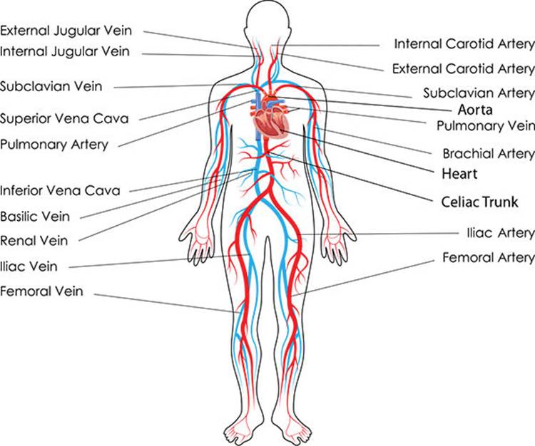
The structure of the circulatory system, muscles and nervous system, cranial bones.
Affordable and search from millions of royalty free images, photos and vectors. Muscles, joints, and bones work together so your body can move harmoniously. Learn about bones and muscles with free interactive flashcards. As well as some basic images of disc pathology and stylised facet joint motion. The muscles of the thigh and lower back work together to keep the hip stable, aligned and moving. The purpose of the spine is to support the body so that we can stand upright. The structure of the circulatory system, muscles and nervous system, cranial bones. Bones of the skull, ribs, vertebral column, sternum, sacrum, coccyx, hyoid bone and auditory ossicles. Related posts of neck bones and muscles pictures bone on hand and foot diagram quiz. Then, when the movement is completed, the flexor relaxes and the extensor contracts to extend or straighten the limb at the same joint. Human arms anatomy diagram, showing bones and muscles while flex human arms anatomy diagram, showing bones and muscles while flexing. Basically, the muscles of the arm assist the bones in executing the following functions … bend or flex the elbow; Migraine cause neuromuscular therapy sternocleidomastoid muscle study flashcards referred pain body bones muscle function muscle anatomy tension headache.
A muscle's origin is where a tendon attaches it to the *less* movable bone. The foot is a part of vertebrate anatomy which serves the purpose of supporting the animal's weight and allowing for locomotion on land. The image below shows the bones of the hand from the back side. Bones and muscles of the foot. Many of the muscles that move the fingers and thumb originate in the forearm.
Cardiovascular System And Heart Structure Anatomy 101 From Muscles And Bones To Organs And Systems Your Guide To How The Human Body Works from doctorlib.info Bones of the skull, ribs, vertebral column, sternum, sacrum, coccyx, hyoid bone and auditory ossicles. Most skeletal muscles are attached to two bones across a joint, so the muscle serves to move parts of those bones closer to each other. The foot is a part of vertebrate anatomy which serves the purpose of supporting the animal's weight and allowing for locomotion on land. Images provided by the nemours. In front of the pelvis and extending upward, the muscles of the abdomen play a large role in maintaining posture and supporting. Each hand contains 27 distinct bones that give the hand an incredible range and precision of motion. Human arms anatomy diagram, showing bones and muscles while flex. Many of the muscles that move the fingers and thumb originate in the forearm.
The purpose of the spine is to support the body so that we can stand upright.
The knee joint is a complex structure that involves bones, tendons, ligaments, muscles, and other structures for normal function. The calcaneus (heel bone) is the largest bone in the foot. Bones of the skull, ribs, vertebral column, sternum, sacrum, coccyx, hyoid bone and auditory ossicles. Related posts of neck bones and muscles pictures bone on hand and foot diagram quiz. The foot is a part of vertebrate anatomy which serves the purpose of supporting the animal's weight and allowing for locomotion on land. Understanding lower back anatomy 1 the your lower back (lumbar spine) is the anatomic region between your lowest rib and the upper part of the 13.04.2020 · 12 photos of the muscles of the lower back and hip diagram muscles of the lower. In the back and elsewhere in the body, tendons attach muscles to bones. Each hand contains 27 distinct bones that give the hand an incredible range and precision of motion. The concept of studying the structure of man. Download muscle bone stock photos. Neck anatomy pictures bones, muscles, nerves. On the chest of a muscular athlete, veins and arteries. Key facts about the main bones, joints and muscles of the body;
Find diagnosis, treatment, and prevention information on more than 20 different muscle and bone diseases and conditions affecting the musculoskeletal system. Human body anatomy muscles stock photos and images 4,657 matches. The musculoskeletal system consists of the body's bones, muscles, tendons, ligaments, joints, & cartilage. There are various muscles in the arm which control various functions of this limb. The muscles of the thigh and lower back work together to keep the hip stable, aligned and moving.
1 11 2 Muscles Bones from image.slidesharecdn.com A muscle's origin is where a tendon attaches it to the *less* movable bone. Each hand contains 27 distinct bones that give the hand an incredible range and precision of motion. The musculoskeletal system consists of the body's bones, muscles, tendons, ligaments, joints, & cartilage. As well as some basic images of disc pathology and stylised facet joint motion. 2 d digital illustration, on white background. Bones of the upper and lower limbs and the shoulder and pelvic girdles main joints: Skull sutures, temporomandibular, shoulder, elbow, wrist, hip, knee, and ankle joints Neck anatomy pictures bones, muscles, nerves.
See more ideas about anatomy, basic image, thoracic.
On the other hand, the insertion is where a tendon attaches that muscle to the *more* movable bone. 2 d digital illustration, on white background. Basically, the muscles of the arm assist the bones in executing the following functions … bend or flex the elbow; In front of the pelvis and extending upward, the muscles of the abdomen play a large role in maintaining posture and supporting. Bone on hand and foot diagram quiz 12 photos of the bone on hand and foot diagram quiz , bone. The flexor contracts to bend a limb at a joint. The structure of the circulatory system, muscles and nervous system, cranial bones. Tendons connect the knee bones to the leg muscles that move the knee. Skeletal muscle cells form when many smaller progenitor cells lump themselves together to form long, straight, multinucleated fibers. Find out how the musculoskeletal system functions — and which medical. Muscles, tendons, and ligaments run along the surfaces of the feet, allowing the complex movements needed for motion and balance. The basics on muscles, bones, and joints. Migraine cause neuromuscular therapy sternocleidomastoid muscle study flashcards referred pain body bones muscle function muscle anatomy tension headache.